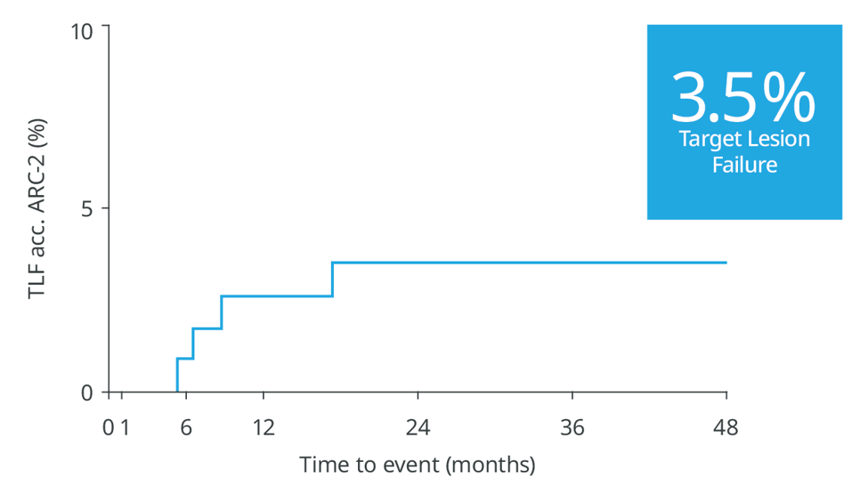
Skip to content

Freesolve

Resorbable Magnesium Scaffold

Metallic performance1-3.
Fully resorbablea,4.

Download the Brochure

Thank you for your interest in Freesolve™ RMS. Please provide your contact details to download the product brochure.

Key Benefits

Delivers like a DES5

Optimal vessel support6,7

Magnesium fully resorbed after 12 months8

Excellent safety and efficacy2,3

Full System

Pre-procedure

Initial diagnostic

Post-procedure

Immediately after Implantation, struts are well apposed to the vessels wall.

6-month follow-up

While the Magnesium resorption process continues, endothelialization progresses.

12-month follow-up

The Magnesium resorption is completed. No struts appear in OCT.

>99%

of struts no longer visible at 12 months8

Magnesium fully resorbed after 12 months8

Procedure

Experience shows that following 4 Ps for Freesolve™ RMS implantations may lead to better patient outcomes.

Patient Selection

Appropriate patient selection is crucial to achieve procedural success. Freesolve™ is currently indicated for de novo lesions, with a reference vessel diameter and lesion length closely matching the available Freesolve™ sizes. Each individual patient should receive best clinical care and should benefit from BRS Technology.

Proper Sizing

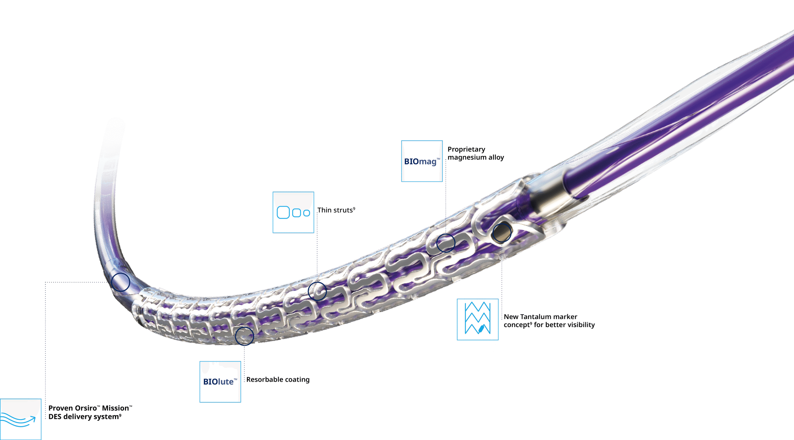
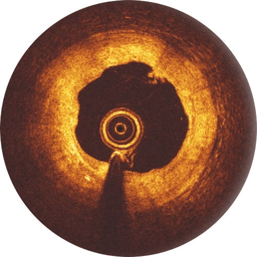
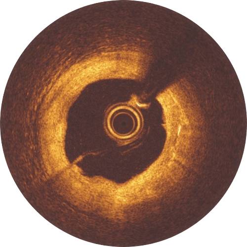
If uncertain about the vessel diameter, use QCA, IVUS and/or OCT for quantitative lesion evaluation. The diameters available are 2.5, 3.0, 3.5 and 4.0 mm, do not implant into vessels 4.2 mm in diameter. Angiogram generally underestimates the diameter of the vessel by 0.25 mm.

Pre-Dilatation

Pre-dilatation with a non-compliant balloon with a 1:1 balloon-to-artery ratio is mandatory. The balloon should expand fully. The residual stenosis before the Freesolve™ implantation is recommended to be less than 20 %. If the pre-dilatation goal is not achieved, use other balloon technologies such as scoring balloons.
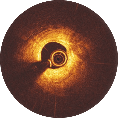
Post-Dilatation

Post-dilatation with a non-compliant balloon 0.5 mm larger than the implanted scaffold expanded at high pressure (> 16 atm) is recommended. Please keep in mind that the Freesolve™ expansion limit is 0.6 mm beyond nominal scaffold size. During the learning phase, OCT is helpful to check for vessel and lumen dimensions, lesion length and struts’ malapposition.

Clinical Highlights

A wealth of clinical evidence

Study nameStudy typePatients in clinical studyTest model in pre-clinical studyStatusEndpointFlyer (where available)
BIOMAG-IIRCT1,859-InitiatedTLF at 12 months
RMS RegistryRegistry1,106-InitiatedTLF at 12 months
BIOMAG-I
FIH 115 - 36m FUP availableIn-scaffold LLL at 6 monthsDownload
BIOMAG-I OCT Analysis* FIH 69 - 12m FUP availableVisible struts at 6 and 12 months
BIOMAG-I Return of vasomotion* FIH 14 - - Vasomotion assessment after Acetylcholine (Ach) and Nitroglycerine (NTG)
BIOMAG-I OCT Plaque characterization (AI-based OCT)* FIH 84/6m 46/12m - - Comparison of different plaque characteristicsDownload
BIOMAG-I Plaque characterization* FIH 84 - - Impact of plaque characteristics on in-stent LLL
BIOSOLVE-IV Full COHORT conducted with Magmaris Registry 2,066 - 36m FUP available Target Lesion Failure* at 12 months
Evaluation of the degradation kinetics*Animal study-mini swineCompleted Degradation kinetics analysis: 99.6% backbone degradation for Freesolve™ at 12 months
Evaluation of Thrombogenicity Using Human BloodIn-vitro study - flow chamber study Completed BIOmag™ scaffold material shows strongly reduced in vitro thrombogenicity
Preclinical animal safety and device characterization up to 4yAnimal study -mini swineOngoingSafety evaluation
Chronic Scaffolding Evaluation for DREAMS 3GIn Silicon study- simulation Ongoing Resorption and Finite element analysis

*restricted access, sign-in required

36-month outcomes confirm: No new events between 2 and 3 years9

 3.5%

Target Lesion Failure

0.0%

Scaffold Thrombosis

0.0%

Myocardial Infarction

0.0%

Cardiac Death